
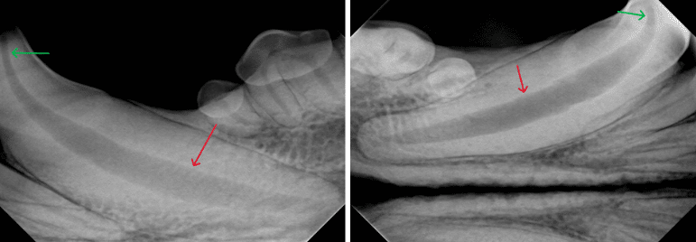
(1a, 1b) Pulpitis in canine mandibular canines; inflammation of the pulp cavity can cause it to widen. In these views, you can see how much the pulp cavity expands in width from the green arrows to the red arrows.
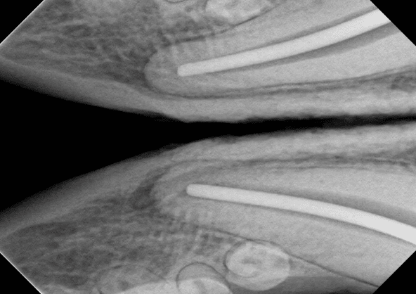
(1c) The same mandibular canines radiographed directly after a root canal.
***All pathology seen on dental radiographs must be considered with the context of a dental examination, and vice versa. The severity of periodontal disease cannot be fully appreciated without looking at the underlying roots and bone structures that make up the maxilla (or upper part of the mouth) and the mandible (the jaw.) The parts of the teeth we see are very much only “the tip of the iceberg.”

(3) Canine maxillary incisors; the red arrow marks where the crown of the innermost incisor on the dog’s left side was broken off. The soft tissue of the gumline is not as easily visible here, and upon visual examination of the mouth this could have just looked like a missing tooth. The x-ray shows that the root of the tooth is still present, and will need to be removed.
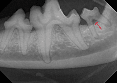
(4) Canine mandibular molar; the red arrow in this view points to where the underlying bone has receded from the tooth, exposing more of the roots. You can see from the neighboring teeth where the bone level should be.

(5) Canine mandibular molar; this view from a small breed dog shows us just how important it is to keep up with dental health. As you can see, the root indicated by the red arrow is much larger than the one with the green arrow, and it extends deeper in the mouth and comes close to the edge of the patient’s jaw. This is a normal occurrence in many dogs, especially small breeds, but if the tooth becomes diseased it can affect the underlying bone, and the structure of the entire jaw could be compromised.

(6a) Canine maxillary premolar; the red arrows mark a retained deciduous (baby) tooth, and the dotted line to the left of it marks the level of the bone holding it in. You can visualize how little of the tooth is actually attached, and the bone loss has extended to the neighboring tooth which had to be extracted.
(6b) After extraction of these two teeth; the level of the bone is more easily visualized. Unfortunately the premolar on the other side hasn’t escaped unscathed; the red arrow marks where the root has been resorbed. That tooth was also extracted.

(6c) Canine maxillary premolar; the same retained deciduous tooth on the opposite side of the mouth. The surrounding bone is not as severely affected on this side, and the tooth was extracted while the neighboring teeth were able to be saved. (Also: note the rotated premolar to the right of it!)

(7) Feline maxillary canine; the red arrow marks severe inflammation of the bone where a canine tooth used to be. Note that the neighboring incisors are missing. This patient had severe periodontal disease at the time of these x-rays.

(8) (left) Feline mandibular premolar; this lonely little tooth had such severe bone loss that it was almost being completely held in by gum tissue. Periodontal disease is described in patients on a scale of zero to four, and this patient is considered to be at stage four, which is the most severe stage.
(8b) (right) This x-ray shows the same feline mandibular premolars on another patient for comparison; the bone is level and covers the entirety of the root. To the right you can see where one tooth has been extracted.

(10a, 10b) Feline mandibular premolar; the red arrow marks a Feline Odontoclastic Resorptive Lesion (FORL), which generally occurs toward the crown of a cat’s tooth. Also shown is that same view after the tooth was extracted.

(11) Feline mandibular premolar; the red arrow marks where part of the tooth’s roots were retained after the tooth had been previously lost or broken. Retained roots can cause painful infection and cannot be visualized without an x-ray.